Welcome to the Amira-Avizo Software Use Case Gallery
Below you will find a collection of use cases of our 3D data visualization and analysis software. These use cases include scientific publications, articles, papers, posters, presentations or even videos that show how Amira-Avizo Software is used to address various scientific and industrial research topics.
Use the Domain selector to filter by main application area, and use the Search box to enter keywords related to specific topics you are interested in.
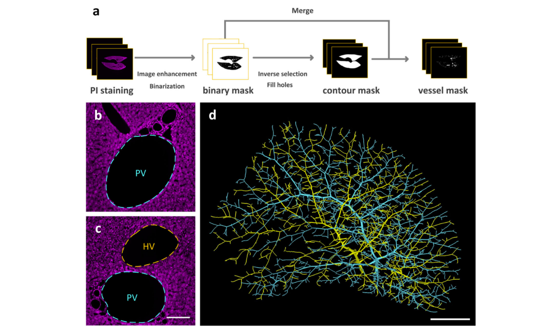
Multiscale reconstruction of various vessels in the intact murine liver lobe
The liver contains a variety of vessels and participates in miscellaneous physiological functions. While past studies generally focused on certain hepatic vessels, we simultaneously obtained all the vessels and cytoarchitectural information of the intact mouse liver lobe at single-cell resolution. […] providing a technology roadmap for studying the fine hepatic vascular structures and their spatial relationship, which will help research into liver diseases and evaluation of medical effi... Read more
Qi Zhang, Anan Li, Siqi Chen, Jing Yuan, Tao Jiang, Xiangning Li, Qingming Luo,Zhao Feng & Hui Gon
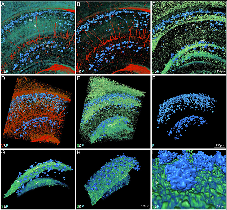
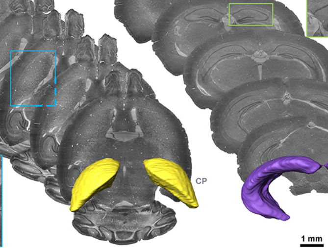
High-Resolution Digital Panorama of Multiple Structures in Whole Brain of Alzheimer's Disease Mice
Our study placed emphasis on solving problems in processing high-throughput bright field images and made attempt in developing a method for the extraction and reconstruction of multiple structures. This will facilitate a better understanding of the cerebral anatomical features under the pathological state of AD and shows extensive application prospect in drug efficacy assessment from brain-wide level.
Simultaneously visualizing Amyloid-β (Aβ) plaque with its surrounding brain structu... Read more
Xianzhen Yin, Xiaochuan Zhang, Jingjing Zhang, Weicheng Yang, Xian Sun, Haiyan Zhang, Zhaobing Gao, Hualiang Jiang
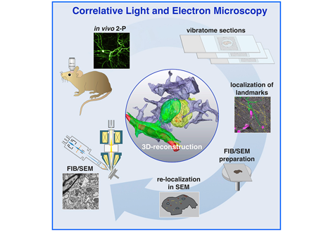
Label-free 3D-CLEM using endogenous tissue landmarks
We demonstrate feasibility of the workflow by combining in vivo 2-photon microscopy and focused ion beam scanning electron microscopy (FIB/SEM) to dissect the role of astrocytic coverage in the persistence of dendritic spines.
Emerging 3D correlative light and electron microscopy (CLEM) approaches enable studying neuronal structure-function relations at unprecedented depth and precision. However, established protocols for the correlation of light and electron micrographs rely ... Read more
Manja Luckner,Steffen Burgold, Severin Filser, Maximilian Scheungrab, Yilmaz Niyaz, Eric Hummel, Gerhard Wanner, Jochen Herms
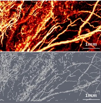
Optical Resolution Photoacoustic Microscopy of Ovary and Fallopian Tube
Ovarian cancer is the leading cause of death among gynecological cancers, but is poorly amenable to preoperative diagnosis. In this study, we investigate the feasibility of “optical biopsy,” using high-optical-resolution photoacoustic microscopy (OR-PAM) to quantify the microvasculature of ovarian and fallopian tube tissue. The technique is demonstrated using excised human ovary and fallopian tube specimens imaged immediately after surgery.
This report describes the first applicatio... Read more
Bin Rao, Xiandong Leng, Yifeng Zeng, Yixiao Lin, Ruimin Chen, Qifa Zhou, Andrea R. Hagemann, Lindsay M. Kuroki, Carolyn K. McCourt, David G. Mutch, Matthew A. Powell, Ian S. Hagemann & Quing Zhu
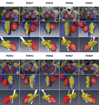
Patient-specific anatomical model for deep brain stimulation based on 7 Tesla MRI
Deep brain stimulation (DBS) requires accurate localization of the anatomical target structure, and the precise placement of the DBS electrode within it. Ultra-high field 7 Tesla (T) MR images can be utilized to create patient-specific anatomical 3D models of the subthalamic nuclei (STN) to enhance pre-surgical DBS targeting as well as post-surgical visualization of the DBS lead position and orientation. We validated the accuracy of the 7T imaging-based patient-specific model of the STN and m... Read more
Yuval Duchin, Reuben R. Shamir, Remi Patriat, Jinyoung Kim, Jerrold L. Vitek, Guillermo Sapiro, Noam Harel
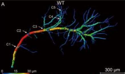
Accumulating evidence indicates the critical importance of cerebrovascular dysfunction in the pathogenesis of Alzheimer’s disease (AD). However, systematic comparative studies on the precise brain vasculature of wild-type and AD model mice are still rare. Using an image optimization method for analyzing Micro-Optical Sectioning Tomography (MOST) data, we generated cross-scale whole-brain 3D atlases that cover the entire vascular system from large vessels down to smallest capillaries at ... Read more
Xiaochuan Zhang, Xianzhen Yin, Jingjing Zhang, Anan Li, Hui Gong, Qingming Luo, Haiyan Zhang, Zhaobing Gao, Hualiang Jiang
Precise Cerebral Vascular Atlas in Stereotaxic Coordinates of Whole Mouse Brain
Understanding amazingly complex brain functions and pathologies requires a complete cerebral vascular atlas in stereotaxic coordinates. Making a precise atlas for cerebral arteries and veins has been a century-old objective in neuroscience and neuropathology. Using micro-optical sectioning tomography (MOST) with a modified Nissl staining method, we acquired five mouse brain data sets containing arteries, veins, and microvessels. Based on the brain-wide vascular spatial structures and brain re... Read more
Benyi Xiong, Anan Li, Yang Lou, Shangbin Chen, Ben Long, Jie Peng, Zhongqin Yang, Tonghui Xu, Xiaoquan Yang, Xiangning Li, Tao Jiang, Qingming Luo and Hui Gong
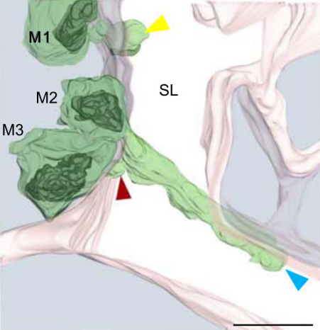
Multiple membrane extrusion sites drive megakaryocyte migration into bone marrow blood vessels
Platelets, cells central to hemostasis and thrombosis, are formed from parent cell megakaryocytes. Although the process is highly efficient in vivo, our ability to generate them in vitro is still remarkably inefficient. We proposed that greater understanding of the process in vivo is needed and used an imaging approach, intravital correlative light electron microscopy, to visualize platelet generation in bone marrow in the living mouse. In contrast to current understanding, we found that most... Read more
Edward Brown, Leo M Carlin, Claus Nerlov, Cristina Lo Celso, Alastair W Poole
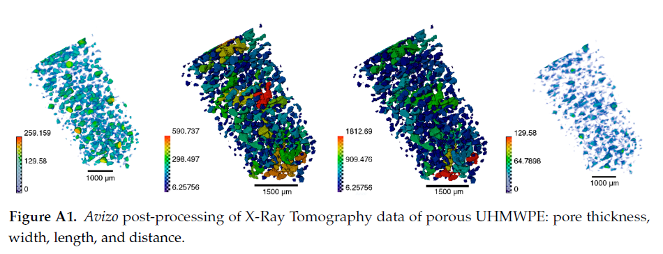
Since its invention and commercialization in the 1950s, ultra-high molecular weight polyethylene (UHMWPE) has been known as a high-performance polymer successfully applied in diverse engineering systems ranging from strong ropes for naval demands and wear-resistant liners in bearings, transportation belts and heavy trucks in mines and quarries, through the lining of chemical vessels and disposable bags in bioreactors, to sophisticated products such as orthopaedic implants and replacements of ... Read more
Eugene S. Statnik, Codrutza Dragu, Cyril Besnard, Alexander J.G. Lunt, Alexey I. Salimon, Aleksey Maksimkin and Alexander M. Korsunsky
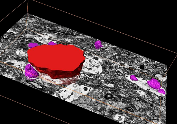
Corpora amylacea are cell-derived structures that appear physiologically in the aged human brain. While their histological identification is straightforward, their ultrastructural composition and microenvironment at the nanoscale have remained unclear so far, as has their relevance to aging and certain disease states that involve the sequestration of toxic cellular metabolites. Here, we apply correlative serial block-face scanning electron microscopy and transmission electron tomograp... Read more
Paula P. Navarro, Christel Genoud, Daniel Castaño-Díez, Alexandra Graff-Meyer, Amanda J. Lewis, Yvonne de Gier, Matthias E. Lauer, Markus Britschgi, Bernd Bohrmann, Stephan Frank, Jürgen Hench, Gabriel Schweighauser, Annemieke J. M. Rozemuller, Wilma D. J. van de Berg, Henning Stahlberg & Sarah H. Shahmoradian